
Knee pain is one of the most common reasons people visit a doctor. It can come from a sudden sports injury, a twist on the stairs, or years of wear on the joint. Whatever the cause, it can limit walking, exercise, work, and sleep.
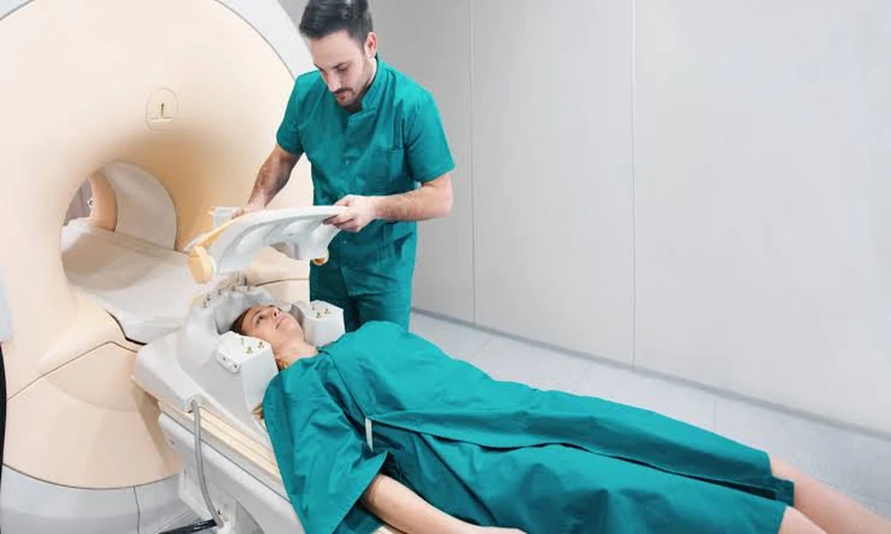
When knee pain does not improve or the cause is unclear, doctors often turn to MRI. This type of imaging gives a detailed look inside the joint and plays a key role in deciding which treatment makes sense. Here is how MRI helps guide care for knee problems.
Why the Knee Is So Vulnerable
The knee is one of the largest and most complex joints in the body. It connects the thighbone to the shinbone and relies on a network of ligaments, tendons, cartilage, and muscles to stay stable. It also carries a large share of your body weight with every step.
That combination of complexity and constant load makes the knee prone to injury. Twisting movements, sudden stops, direct hits, and repetitive stress can all damage different parts of the joint. Because many structures sit close together, the source of pain is not always obvious from a physical exam alone.
When Does Knee Pain Call for an MRI?
Not every sore knee needs advanced imaging. Many minor strains improve with rest, ice, and time. Doctors usually start with a physical exam and sometimes an X-ray, which is useful for showing bones and signs of arthritis.
An MRI may be recommended when pain persists for weeks, when the knee feels unstable or gives way, or when it locks or catches during movement. Significant swelling after an injury is another common reason. MRI is also helpful when symptoms do not match what an X-ray shows.
What an MRI Can Reveal in the Knee
Unlike X-rays, MRI shows soft tissues in great detail. This makes it especially valuable for the knee, where many important structures are not bone. MRI uses magnetic fields and radio waves, not radiation, to create its images.
Ligament injuries are one of the most common findings. The anterior cruciate ligament, or ACL, and the medial collateral ligament, or MCL, are frequently injured in sports. MRI can show whether a ligament is stretched, partially torn, or fully torn.
Meniscus tears are another frequent problem. The meniscus is a C-shaped cushion of cartilage that absorbs shock between the bones. MRI can show the location, size, and pattern of a tear, which strongly influences treatment.
MRI can also reveal cartilage damage, bone bruises, tendon injuries, fluid in the joint, and early signs of arthritis that may not appear on X-rays yet.
How MRI Findings Shape Treatment Decisions
The details from an MRI help doctors match the treatment to the actual problem. Two people with similar knee pain may need very different care depending on what the images show.
For example, a small meniscus tear on the outer edge, where blood supply is better, may heal or be managed with physical therapy. A larger tear that causes locking may need arthroscopic surgery to repair or trim it. In the same way, a partial ligament strain often heals with bracing and rehab, while a complete ACL tear in an active person may lead to a discussion about reconstruction.
Clear imaging also helps doctors avoid unnecessary procedures. If the MRI shows no structural damage that needs surgery, the focus can shift to strengthening, mobility work, and other conservative treatments.
The Value of Weight-Bearing Knee Imaging
Traditional MRI scans the knee while the patient lies down with the leg relaxed. This works well for many conditions. However, the knee behaves differently when it supports body weight, as it does when standing or walking.
Some open MRI systems allow the knee to be imaged while the patient stands or sits in a weight-bearing position. Through upright MRI in Deerfield, for example, patients can be scanned in positions closer to how the knee works in daily life. This may give doctors additional insight into joint alignment and how structures respond under load.
Comfort Matters During a Knee Scan
Staying still is important for clear MRI images. Patients with significant knee pain may struggle to hold one position for long, and those who feel anxious in closed scanners may find the experience stressful.
Open MRI designs can help. They allow patients to see their surroundings and often feel less confined. A more comfortable patient is usually able to stay still, which leads to better image quality and fewer repeat scans.
Preparing for Your Knee MRI
Preparation for a knee MRI is usually simple. You will be asked to remove metal objects such as jewelry, watches, and belts. Let the imaging team know about any implants, pacemakers, or metal in your body before the scan.
Wear comfortable clothing without metal zippers or snaps if possible. Bring your doctor’s order, insurance details, and any previous imaging reports. Asking how long the scan will take and when results will reach your doctor can help you plan.
Conclusion
MRI gives doctors a clear look at the ligaments, cartilage, tendons, and bones inside the knee. These details help them understand the true cause of pain and choose between rest, physical therapy, injections, or surgery with greater confidence.
If your doctor recommends imaging, ask whether weight-bearing scanning might add useful information for your case. Physicians can also learn more about referring patients for positional MRI, helping knee patients get imaging that supports the right treatment plan.